
Our brain is an incredible electrical powerhouse, where billions of neurons constantly send signals to coordinate everything we feel, think, and do. But how can scientists actually "listen" to this silent electrical symphony? With sophisticated electrophysiology and brain imaging tools, researchers can now track, map, and even influence these signals, giving us a glimpse into the brain's inner workings.
Whether through single-neuron recording, MRI imaging, or techniques like PET scans, each method brings us one step closer to understanding the mysteries behind our thoughts and emotions. Here’s a look into the incredible techniques that make this possible.
Single-Neuron Recording: Tuning In on Individual Brain Cells
Imagine zooming in on a single neuron and tracking every time it fires. That’s exactly what single-neuron recording achieves, allowing scientists to capture the real-time activity of one neuron as it communicates with others.
- How It Works: This technique places tiny electrodes near a single neuron to pick up every action potential—or electric signal—it sends. With this setup, researchers can monitor neuron activity moment by moment.
- Why It Matters: Single-neuron recording has helped reveal how certain neurons respond to specific tasks or sights. For instance, some neurons activate in response to a familiar face, while others fire as an animal explores a new space. These insights help us understand individual neuron roles in perception, memory, and behavior.
Patch-Clamp Recording: Exploring Ion Channels
Ever wondered how neurons send signals across the brain? The patch-clamp technique lets scientists observe the tiny ion channels that control these signals.
- How It Works: A glass pipette attaches to a small part of the neuron’s cell membrane, allowing researchers to record the ion flow in and out of specific channels. By controlling the environment, they can see how channels open and close in response to changes.

Attribution: Pavel Novak, Julia Gorelik, Umesh Vivekananda, Andrew I. Shevchuk, Yaroslav S. Ermolyuk, Russell J. Bailey, Andrew J. Bushby, Guy W.J. Moss, Dmitri A. Rusakov, David Klenerman, Dimitri M. Kullmann, Kirill E. Volynskicorrespondenceemail, Yuri E. Korchev, CC BY 3.0 <https://creativecommons.org/licenses/by/3.0>, via Wikimedia Commons 
Attribution: Gregory Holst, CC BY-SA 3.0 <https://creativecommons.org/licenses/by-sa/3.0>, via Wikimedia Commons
- Why It Matters: Patch-clamp recording has been invaluable in neuroscience, giving insight into cellular communication at the most fundamental level. It’s a critical tool for testing drugs that target ion channels, which are key players in treating neurological diseases.
ECG: Tracking the Heart’s Electrical Signals
Though the brain often steals the spotlight, the heart has its own powerful rhythm driven by electrical signals. The electrocardiogram (ECG) is a simple, non-invasive way to measure these signals.
 |
| Attribution: Created by Agateller (Anthony Atkielski), converted to svg by atom., Public domain, via Wikimedia Commons |
- How It Works: Electrodes are placed on the skin, tracking the heart’s rhythm as it pumps blood. The data produces waveforms that display the heart’s cycle of contraction and relaxation.
- Why It Matters: ECGs help doctors diagnose heart conditions like arrhythmias or even heart attacks. Researchers also use ECGs to explore how factors like stress, exercise, and medication influence heart health.
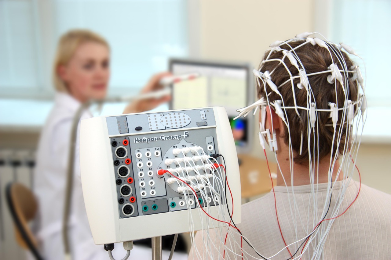
EEG and MEG: Measuring Brainwaves in Real-Time
The brain generates not just individual signals but also waves of electrical activity, visible with EEG and MEG. These waves reveal insights into different mental states and brain functions.
- Electroencephalography (EEG): EEG uses electrodes on the scalp to measure the brain’s electrical activity, creating patterns that align with states like sleep, relaxation, and focus.
 |
| Attribution: Baburov, CC BY-SA 4.0 <https://creativecommons.org/licenses/by-sa/4.0>, via Wikimedia CommonsBaburov, CC BY-SA 4.0 <https://creativecommons.org/licenses/by-sa/4.0>, via Wikimedia Commons |
- Magnetoencephalography (MEG): MEG tracks magnetic fields generated by the brain’s electric currents, showing where the activity is coming from. MEG is often used in epilepsy studies and brain mapping.
 |
| Attribution- Unknown NIMH author, Public domain, via Wikimedia Commons |
- Applications: EEG and MEG are crucial in understanding epilepsy, sleep, and cognition. These tools capture brain responses in real time, making them valuable for studying how we process information and emotions.
Lesion and Stimulation Techniques: Exploring Brain Function Through Intervention
Understanding the brain sometimes means disrupting its processes to see what changes. Lesion and stimulation techniques help scientists study specific brain areas and their impact on behavior.
- Lesions: By studying natural lesions (or injuries) in specific brain areas, scientists can see the roles these regions play. Although we don’t induce lesions in humans, naturally occurring brain injuries provide critical insights.
- Stimulation: Deep Brain Stimulation (DBS) and Transcranial Magnetic Stimulation (TMS) activate certain areas, allowing researchers to observe effects on mood, movement, or memory. DBS is even used to reduce symptoms in Parkinson’s disease patients.
- Why It Matters: These approaches have transformed our understanding of brain functions, especially regarding motor control, memory, and emotional regulation. They are also invaluable for developing new treatments.
Pharmacological Testing: Observing the Brain’s Reaction to Drugs
Studying how drugs affect the brain is a key way to explore its functions. Pharmacological testing uses specific compounds to see how brain activity changes, revealing insights into neurotransmitters and receptors.
- How It Works: By administering drugs that affect certain receptors, researchers observe changes in brain function and behavior, tracing the pathways and mechanisms involved.
- Applications: This method is used to develop treatments for conditions like depression, schizophrenia, and Parkinson’s disease. By observing how neurotransmitters like dopamine influence behavior, scientists better understand their roles in mood, reward, and cognition.
Brain Imaging: PET, MRI, fMRI, and CAT Scans
For a closer look at the brain, advanced imaging techniques like PET, MRI, fMRI, and CAT are essential. These scans reveal the brain’s structure and activity, each with unique advantages.
PET (Positron Emission Tomography)
- How It Works: PET uses a radioactive tracer that binds to certain brain molecules. The tracer’s signals show areas of high metabolic activity.
 |
| Attribution: Jens Maus (http://jens-maus.de/), Public domain, via Wikimedia Commons |
- Applications: PET helps researchers study conditions like Alzheimer’s disease by tracking metabolic changes, giving insights into brain chemistry and disease progression.
MRI (Magnetic Resonance Imaging)
- Functionality: MRI captures detailed images using magnetic fields, ideal for viewing the brain’s structure and distinguishing between gray and white matter.
 |
| Attribution: Shivani Mattikalli/The Quantum Atlas, CC BY-SA 4.0 <https://creativecommons.org/licenses/by-sa/4.0>, via Wikimedia Commons |
- Applications: MRI is a staple for diagnosing structural issues like tumors, strokes, or lesions, giving doctors a highly detailed look at the brain’s anatomy.
fMRI (Functional MRI)
- How It Works: By measuring blood flow changes, fMRI identifies active brain regions during tasks or thoughts, mapping cognitive functions in detail.
 |
| Attribution- Washington irving at English Wikipedia, Public domain, via Wikimedia Commons |
- Applications: fMRI is widely used to study language, memory, and emotions, allowing researchers to map the brain’s responses to different cognitive and emotional stimuli.
CAT (Computed Axial Tomography)
- Functionality: CAT (or CT scanning) uses X-rays to create cross-sectional brain images, especially useful for identifying injuries and bleeding.
 |
| Attribution- Mikael Häggström, CC0, via Wikimedia Commons |
- Clinical Use: CAT is often the first choice in emergency settings for diagnosing traumatic brain injuries, stroke, and hemorrhages.
The Future of Brain Research: Combining Techniques for Deeper Insights
While each technique shines on its own, combining them allows for a fuller view of brain function. By merging electrophysiology, pharmacological interventions, and imaging, researchers can study complex circuits and brain areas in detail. This synergy is opening new doors in brain-computer interfaces, neuroprosthetics, and mental health treatments, promising innovative ways to address neurological conditions.
Conclusion: Unlocking the Brain’s Electric Language
Through electrophysiology and imaging, scientists can now listen to the brain’s electric language, uncovering secrets about perception, memory, and consciousness. These techniques allow us not only to understand the brain but also to find new ways to treat and even enhance it, bridging the gap between mystery and understanding.