
Imagine if we could zoom in on life itself, seeing not just cells but even the bustling structures within them. Microscopy has made this possible, revolutionizing science and medicine. From the earliest light microscopes to cutting-edge electron microscopes, we can now observe the incredible activities that shape life at the tiniest scales.
Join us on a journey through the different types of microscopes and the fascinating techniques that let scientists visualize, understand, and appreciate the microscopic mysteries of life.
A Window into Cells: The Evolution of Light Microscopy
Light microscopy was where it all began. The concept is straightforward—light is used to magnify small objects, giving us a closer look at cellular structures. But how close? With light microscopes, we can reach a resolution of around 200 nanometers (nm). This lets us see structures like the cell nucleus and some larger organelles, but we hit a wall when it comes to finer components like ribosomes or proteins.
_-_larger_text.png) |
| Attribution- Mikael Häggström, M.D. Author info - Reusing images- Conflicts of interest: NoneMikael Häggström, M.D., CC0, via Wikimedia Commons |
 |
| Attribution: FDominec, CC BY-SA 4.0 <https://creativecommons.org/licenses/by-sa/4.0>, via Wikimedia Commons |
Getting More Detail: Fluorescent and Confocal Microscopy
By adding fluorescent dyes and proteins that light up under special conditions, we can pick out particular molecules, almost like highlighting them. Confocal microscopy then uses lasers to build crisp, detailed 3D images. Together, these techniques allow us to see individual parts of cells and track changes over time.
 |
This photograph is of a sample of white-tailed deer sperm stained with peanut agglutinin viewed under fluorescent microscopy at 400x. |
.jpg) |
| Attribution: ZEISS Microscopy from Germany, CC BY-SA 2.0 <https://creativecommons.org/licenses/by-sa/2.0>, via Wikimedia Commons |
Limits of Light
While light microscopes are immensely valuable, they’re still limited by the wavelength of light itself. This led scientists to explore a new frontier—electron microscopy—for deeper clarity.
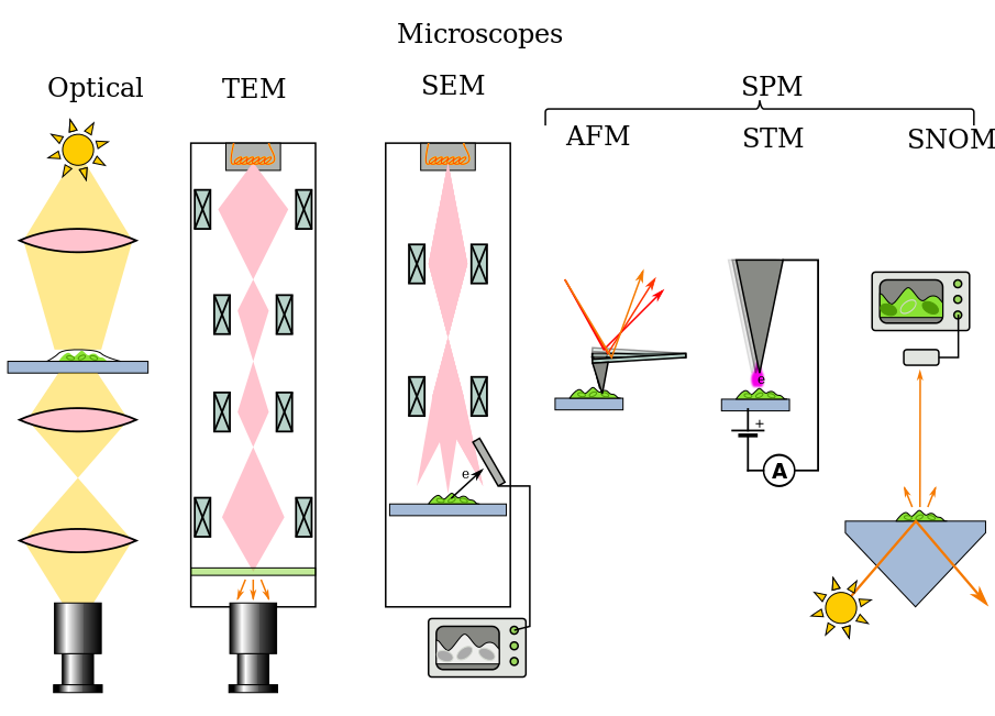
 |
| Attribution: Gringer, CC BY-SA 3.0 <http://creativecommons.org/licenses/by-sa/3.0/>, via Wikimedia Commons |
 |
| Attribution: Leeds University Library, CC BY-SA 4.0 <https://creativecommons.org/licenses/by-sa/4.0>, via Wikimedia Commons |
Microscopy of Living Cells: Observing Life Unfold
To truly understand cellular life, scientists needed a way to study living cells without disrupting their natural state.
Techniques for Visualizing Live Cells
Techniques like Phase Contrast and Differential Interference Contrast (DIC) make it possible to view cells that are transparent and unstained, showing fine details while keeping the cells alive. Another technique, live-cell imaging, uses fluorescent proteins like GFP (Green Fluorescent Protein) to mark specific cellular components. This allows scientists to track molecules as they interact and move, revealing processes like cell division and organelle movement in real time.
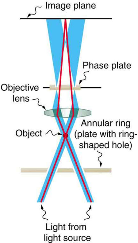 |
| Fig.- Simplified construction of phase contrast microscope Attribution: Connexions, CC BY 3.0 <https://creativecommons.org/licenses/by/3.0>, via Wikimedia Commons |
 |
| Fig.- Diagram illustrating the path of light through a differential interference contrast microscope. Attribution: DIC_Light_Path.png: Richard Wheeler (Zephyris)derivative work: Cepheiden, CC BY-SA 3.0 <http://creativecommons.org/licenses/by-sa/3.0/>, via Wikimedia Commons |
Keeping Cells Happy
Live-cell microscopy requires precise temperature and environmental controls. Cells are sensitive, so scientists must create the right conditions to observe natural behaviors without interfering.
Breaking New Ground: Scanning and Transmission Electron Microscopy
For the most intricate details, electron microscopes (EM) use electrons instead of photons, bringing the resolution down to the nanometer scale and beyond.
Going Deep with Transmission Electron Microscopy (TEM)
Transmission Electron Microscopy (TEM) sends electrons through thin slices of samples, capturing exquisite details within cells down to 0.1 nm. With TEM, scientists can see the internal workings of cells, such as membrane-bound organelles and viral particles, which is especially useful for molecular biology and virology.
.jpg) |
| Attribution: NIAID, CC BY 2.0 <https://creativecommons.org/licenses/by/2.0>, via Wikimedia Commons |
 |
| Attribution: Black Tubus, CC BY-SA 4.0 <https://creativecommons.org/licenses/by-sa/4.0>, via Wikimedia Commons |
 |
Fig.- Transmission electron microscopy observation of “''Ca.'' Midichloria mitochondrii” bacteria in ''Ixodes ricinus'' oocytes Attribution: Francesco Comandatore, Giacomo Radaelli, Sebastiano Montante, Luciano Sacchi, Emanuela Clementi, Sara Epis, Alessandra Cafiso, Valentina Serra, Massimo Pajoro, Domenico Di Carlo, Anna Maria Floriano, Fabrizia Stavru, Claudio Bandi, Davide Sassera, CC BY 4.0 <https://creativecommons.org/licenses/by/4.0>, via Wikimedia Commons |
Exploring Surfaces with Scanning Electron Microscopy (SEM)
Scanning Electron Microscopy (SEM), on the other hand, focuses on scanning the surface of samples with electron beams, creating high-resolution 3D images. SEM is ideal for viewing surface textures and structures—like the delicate shapes of cellular membranes or the intricate details of tissue architecture.
.jpg) |
| Attribution: ZEISS Microscopy from Germany, CC BY-SA 2.0 <https://creativecommons.org/licenses/by-sa/2.0>, via Wikimedia Commons |
_01.jpg) |
| Attribution: Tadeáš Bednarz, CC BY-SA 4.0 <https://creativecommons.org/licenses/by-sa/4.0>, via Wikimedia Commons |
| Attribution: Freundchen, CC0, via Wikimedia Commons |
.jpg) |
| Attribution: ZEISS Microscopy from Germany, CC BY-SA 2.0 <https://creativecommons.org/licenses/by-sa/2.0>, via Wikimedia Commons |
Freeze-Fracture and Freeze-Etch: Peeling Back Layers in Electron Microscopy
Studying delicate samples in their natural state can be challenging. Traditional methods like chemical fixation and sectioning may create artifacts or distortions. Techniques like freeze-fracture and freeze-etch are different; they freeze cells rapidly and then split them along natural fracture lines, preserving cellular integrity.
- Freeze-fracture exposes cell interiors, showing us membranes and other internal structures.
.jpg) |
| Attribution: Wolfbenjamin25, CC BY-SA 4.0 <https://creativecommons.org/licenses/by-sa/4.0>, via Wikimedia Commons |
- Freeze-Etch sublimates ice from the sample’s surface after freeze-fracturing, creating clearer contrasts. This method reveals the true shape of cells without chemical interference, especially useful for observing complex membrane structures.
Fixation and Staining Techniques in Electron Microscopy: Setting the Scene
Preparing samples for electron microscopy is an art of its own. Since electrons need precise conditions to reveal details, proper fixation and staining are crucial.
- Chemical Fixation: Chemicals like glutaraldehyde and osmium tetroxide stabilize the cellular structure by cross-linking proteins.
- Staining with Heavy Metals: Elements like uranium and lead scatter electrons, enhancing the contrast in EM images and making molecular details easier to detect.
- Cryo-EM: A game-changer in sample preparation that freezes samples quickly without fixatives or stains, preserving samples at near-atomic resolution. Cryo-EM has been vital for understanding complex protein structures and molecular assemblies.
Image Processing in Microscopy: Revealing New Depths with Digital Tools
Modern microscopy is enhanced by powerful image processing techniques, allowing scientists to refine, analyze, and extract more information from their observations.
- Deconvolution: This removes out-of-focus light from images, providing clearer, more detailed 3D structures.
- Super-Resolution Imaging: Techniques like STORM (Stochastic Optical Reconstruction Microscopy) and PALM (Photoactivated Localization Microscopy) go beyond the limits of traditional microscopy, achieving resolution comparable to that of electron microscopes.
- AI and Machine Learning: AI-based tools analyze patterns in microscopy data, identifying cell structures, counting cells, and even predicting molecular interactions from observed patterns.
Microscopy’s Impact Across Science and Medicine
Microscopy doesn’t just benefit biology; it plays a central role in medicine, materials science, and even drug discovery.
- Medical Diagnostics: Pathologists use microscopy to spot abnormal cells, bacteria, and viruses, crucial for diagnosing diseases.
- Material Science: Electron microscopy has become essential in developing stronger, lighter, and more durable materials, helping industries innovate and improve products.
- Drug Development: Microscopy aids in drug testing by allowing scientists to see how cells respond to potential treatments, streamlining the development process.
Conclusion: A Clearer Future in Microscopy
Microscopy has transformed science, offering an unparalleled look into the building blocks of life. The advances in super-resolution techniques, improved sample preparation, and AI-driven image processing promise to expand our understanding of the microscopic world further. Each layer we peel back, each new technique we develop, reveals that there’s always more to see and more to discover.