
Radiolabeling might sound like something out of a spy novel, but it’s actually one of the most practical and impactful tools in modern biology. By adding radioactive tags to specific molecules, scientists can track their movement and understand how they work within cells. This technique gives researchers a unique way to look inside living organisms and see molecular processes in real time. Imagine being able to follow a molecule of DNA as it gets copied in a dividing cell or watching how nutrients get absorbed in real time—this is what radiolabeling allows us to do.
In this blog, we’re going to explore everything that makes radiolabeling so special. We’ll dive into the different types of radioisotopes commonly used, break down how scientists incorporate these isotopes into biological cells and look at some of the advanced imaging techniques used to visualize these processes. Finally, we’ll discuss the essential safety measures required when handling radioactive materials. Let’s shed some light on this illuminating technique!
 |
| Fig.- Ball-and-stick model of the component ions in sodium pertechnetate molecule, a radioactive tracer compound used in medical imaging. Attribution-Jynto, CC0, via Wikimedia Commons |
What is Radiolabeling, and Why Do We Use It?
Radiolabeling is all about adding radioactive isotopes to molecules. These isotopes emit a form of radiation that we can detect, so scientists can watch where they go and how they interact with other molecules in a living system. This technique is essential because it gives us a way to understand what’s happening inside cells without interrupting natural processes.
Why Are Radioactive Tracers So Useful?
- Incredible Sensitivity: Thanks to the clear signal emitted by radioactive tracers, even tiny quantities of a molecule can be detected.
- Quantitative Power: Radiolabeling allows for exact measurements, which are especially valuable in biological research.
- Real-Time Observation: Researchers can monitor what’s happening inside cells as it happens, giving dynamic insights into cellular activities.
Types of Radioisotopes Used in Biology
Each isotope has unique properties, from how long it lasts (half-life) to the energy it releases. These properties make certain isotopes more effective for particular studies. Here’s a look at some of the most commonly used radioisotopes in biology:
- Tritium (^3H): Low-energy beta emissions make it safe and great for metabolic studies.
- Carbon-14 (^14C): Its long half-life is ideal for tracking organic molecules over extended periods.
- Phosphorus-32 (^32P): Common in DNA studies due to its strong beta emissions.
- Sulfur-35 (^35S): This beta-emitter works well for tracking proteins.
- Iodine-125 (^125I): Used in immunoassays and binding studies for precise tracking in small protein studies.
How Do We Get Radioisotopes Into Cells?
Incorporating radioisotopes into biological cells and tissues allows scientists to see exactly how molecules interact in living systems. But this incorporation isn’t as simple as it sounds! Different techniques are used depending on what researchers want to track and where they need the radioactive markers to go.
Incorporation Techniques for Biological Research
Direct Labeling: This method involves attaching the isotope straight to the molecule. For example, iodine-125 is commonly used to tag proteins.
Biosynthetic Labeling: Cells are grown in a special medium containing radioactive nutrients, and as they grow, they naturally incorporate these isotopes into their DNA, proteins, or other molecules.
Indirect Labeling: Here, a smaller radioactive molecule is attached to a larger molecule that scientists want to study. This technique is popular in protein studies.
Real-World Applications of Radiolabeling
- Tracking Cell Division: By adding phosphorus-32 to DNA, researchers can monitor how cells replicate.
- Protein Turnover: Using sulfur-35 in amino acids helps researchers study how proteins are broken down and synthesized in cells.
- Lipid Metabolism: Glucose labeled with carbon-14 allows for a deep look into lipid biosynthesis pathways.
How Do Scientists See Radioactive Molecules?
Once molecules are labeled, scientists use imaging techniques to track them in live cells or even entire organisms. These tools have transformed biology by providing a way to look inside living organisms without disrupting their natural processes.
Imaging Techniques for Radioactive Molecules
- Positron Emission Tomography (PET): PET scans use isotopes that release positrons, like fluorine-18, to capture high-resolution images of the brain, tumors, and metabolic activity.
 |
| Attribution- Jens Maus (http://jens-maus.de/), Public domain, via Wikimedia Commons |
- Single Photon Emission Computed Tomography (SPECT): This technique uses single-photon emitters (like iodine-123) and is popular in neurology and cardiology for tracking blood flow.
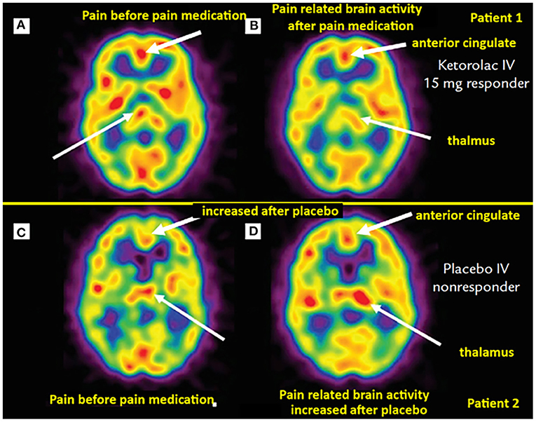 |
| Fig.- Brain perfusion SPECT shows dental pain patients with analgesia (top row) versus placebo (bottom row). Attribution: Newberg et al., CC BY 4.0 <https://creativecommons.org/licenses/by/4.0>, via Wikimedia Commons |
- Autoradiography: For this method, a sample like tissue or cells is placed on film, and radiation exposes the film to create an image.
- Scintillation Counting: By measuring light pulses caused by radiation in a sample, scientists can perform precise quantitative analysis of radiolabeled samples.
Applications of Radioimaging in Biological Research
- Cancer Detection: PET and SPECT scans are crucial in identifying and tracking tumor growth.
- Brain Research: By using tracers, scientists can observe neurotransmitter activity, advancing our understanding of diseases like Parkinson’s.
- Drug Development: Radiolabeling helps track a drug's journey through the body, providing essential data for drug safety and efficacy.
How Do Scientists Measure Radioisotopes in the Lab?
Detecting and measuring the radioisotopes in samples is critical. Different methods are used depending on the type of radiation and the level of sensitivity needed.
Main Detection Techniques
Geiger-Müller Counters: This method is widely used for beta and gamma emissions and provides a quick, straightforward reading of radiation levels.
Liquid Scintillation Counters: For beta emissions, these counters measure light pulses and are perfect for detecting low-energy particles like tritium and carbon-14.
Gamma Counters: Specifically for gamma emissions (like iodine-125), these counters are sensitive and commonly used in medical and biological labs.
Radiographic Films and Phosphor Screens: Used in autoradiography, these methods capture the distribution of radioactivity in samples, allowing scientists to visualize exactly where radiolabeled molecules are within the sample.
Important Aspects of Quantification
- Counting Efficiency: The higher the counting efficiency, the more accurate the measurements are.
- Decay Rate: Each radioisotope has a specific decay rate, and monitoring this is essential for getting reliable measurements over time.
Staying Safe While Working with Radiolabels
Working with radioactive materials means researchers must take strict precautions. These safety measures protect both the scientists and the environment, and they’re mandated by agencies like the NRC and IAEA.
Safety Measures in Radiolabeling Labs
Protective Equipment and Shielding: Lab coats, gloves, and special shields keep exposure levels low. Different materials block different types of radiation—for instance, beta particles are stopped by plastic, but gamma rays require lead.
Radiation Dosimetry: By wearing personal dosimeters, lab workers track exposure levels to ensure they stay within safe limits.
Reducing Exposure Time: Handling samples quickly and efficiently reduces exposure to radiation. Many labs use automated equipment to limit the time spent handling radioactive materials.
Waste Disposal: Radioactive waste must be carefully disposed of according to strict guidelines, often requiring specialized containers and treatment.
Routine Training: Regular training keeps lab workers informed of the latest safety protocols and emergency procedures, helping maintain a safe working environment.
Conclusion: Why Radiolabeling is a Game-Changer in Biology
Radiolabeling continues to be one of the most valuable tools for understanding life on a molecular level. Whether it’s helping scientists watch a cell divide, track a drug through the body, or observe how a tumor responds to treatment, radiolabeling shines a light on some of biology’s greatest mysteries. As technology advances, new types of isotopes and even hybrid imaging techniques (like PET/MRI) promise to expand the horizons of this incredible field even further. By revealing what’s happening at the tiniest levels, radiolabeling paves the way for breakthroughs that could change medicine, environmental science, and our understanding of life itself.