
Imagine being able to zoom into the microscopic world and explore the building blocks of life—molecules—and understand how they work together to make life possible. That’s exactly what biophysical methods allow scientists to do. These powerful techniques provide insights into molecular structures, how molecules interact, and even how they change in real-time, transforming our understanding of biology and chemistry. By using tools like UV/visible spectroscopy, NMR, X-ray diffraction, and mass spectrometry, researchers can uncover details that are invisible to the naked eye but essential to understanding everything from cellular processes to drug design.
In this blog, we’ll walk through some of the most important biophysical methods, how they work, and how they’re helping scientists dive deep into the molecular world. Whether you’re interested in how these methods drive medical research or simply curious about how scientists study molecules, this guide has you covered!
UV/Visible Spectroscopy: Seeing Molecules in Color
UV/visible spectroscopy is a simple yet powerful technique for identifying and analyzing molecules. It’s based on the idea that different molecules absorb light at specific UV or visible wavelengths. When scientists analyze the light a molecule absorbs, they can gather valuable information about its structure, concentration, and even how it interacts with other molecules.
 |
| Attribution: Jon Chui, CC BY-SA 3.0 <https://creativecommons.org/licenses/by-sa/3.0>, via Wikimedia Commons |
How UV/Visible Spectroscopy Works
When light passes through a sample, certain wavelengths are absorbed based on the molecule’s unique structure. A spectrophotometer measures the amount of light absorbed, generating a unique “fingerprint” for each molecule. This absorption pattern reveals a lot about what’s in the sample.
 |
| Attribution- Sobarwiki, Public domain, via Wikimedia Commons |
Applications of UV/Visible Spectroscopy
UV/visible spectroscopy is essential in fields like biology and chemistry. For example, DNA, RNA, and proteins each have unique absorption peaks (like DNA, which absorbs strongly at 260 nm). This technique is also widely used to study reaction rates in enzyme activity and analyze the purity of samples.
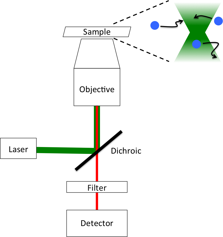
Fluorescence Spectroscopy: Lighting Up Molecular Interactions
Fluorescence spectroscopy allows scientists to “light up” specific molecules and track their interactions. This method takes advantage of molecules that emit light when exposed to certain wavelengths. It’s especially helpful for visualizing molecular interactions within cells or in solution.
 |
| Attribution: Mllyjn, CC BY-SA 3.0 <https://creativecommons.org/licenses/by-sa/3.0>, via Wikimedia Commons |
How Fluorescence Spectroscopy Works
A molecule absorbs light at one wavelength, which excites its electrons to a higher energy state. When the electrons return to their original state, the molecule emits light at a longer wavelength. This emitted light is detected, providing information about molecular interactions and conformational changes.
 |
| Attribution- Sobarwiki, Public domain, via Wikimedia Commons |
Applications of Fluorescence Spectroscopy
Fluorescence spectroscopy is widely used in biology. Researchers often label proteins or other molecules with fluorescent tags, allowing them to track these molecules in living cells. It’s also used to study protein folding and enzyme activities, making it indispensable in cellular and molecular research.
Circular Dichroism (CD): Studying Protein and Nucleic Acid Structure
Circular dichroism (CD) is a specialized technique for examining the secondary structures of proteins and nucleic acids. CD measures the difference in absorption between left- and right-handed circularly polarized light, revealing details about structures like alpha-helices, beta-sheets, and random coils.
How Circular Dichroism Works
When circularly polarized light passes through a chiral molecule, such as a protein or DNA, it absorbs one type of circular polarization more than the other. This difference creates a characteristic CD spectrum that reveals information about the molecule’s secondary structure.
Applications of Circular Dichroism
CD spectroscopy is widely used to analyze protein folding and stability, especially in drug discovery. It helps ensure that drug candidates maintain their structure under different conditions, supporting the development of effective, stable therapeutics.
 |
| Attribution: KingisNitro, CC BY-SA 4.0 <https://creativecommons.org/licenses/by-sa/4.0>, via Wikimedia Commons |
NMR Spectroscopy: Mapping Molecules Atom by Atom
Nuclear Magnetic Resonance (NMR) spectroscopy is one of the most powerful techniques for understanding the three-dimensional structure and dynamics of molecules. By studying how atomic nuclei respond to magnetic fields, NMR can reveal the precise arrangement of atoms within a molecule.
How NMR Spectroscopy Works
NMR relies on the magnetic properties of certain nuclei, such as hydrogen. When these nuclei are placed in a magnetic field and exposed to radiofrequency pulses, they produce signals that reflect their local chemical environment. By analyzing these signals, scientists can map out the positions and connectivity of atoms within the molecule.
| Attribution: Abyss nuko, CC BY-SA 3.0 <https://creativecommons.org/licenses/by-sa/3.0>, via Wikimedia Commons |
Applications of NMR Spectroscopy
NMR is invaluable for determining the structure of complex molecules like proteins, nucleic acids, and small organic compounds. It’s also used to study molecular dynamics, such as changes in shape when molecules bind to other molecules. NMR has become an essential tool in drug development, structural biology, and metabolomics.
Electron Spin Resonance (ESR): Examining Free Radicals and Metals
Electron Spin Resonance (ESR), also known as Electron Paramagnetic Resonance (EPR), is used to study molecules with unpaired electrons, such as free radicals and certain metal ions. ESR provides insight into the electronic structure and the local environment around these molecules.
How ESR Works
When exposed to a magnetic field, unpaired electrons absorb energy at specific frequencies, creating a unique ESR spectrum. This spectrum provides information about the molecular environment, revealing details that are hard to observe with other techniques.
Applications of ESR
ESR is particularly useful for studying free radicals, which are involved in aging, oxidative stress, and diseases like cancer. It’s also used to examine metalloproteins and complex materials, making ESR valuable in biochemistry, environmental science, and materials research.
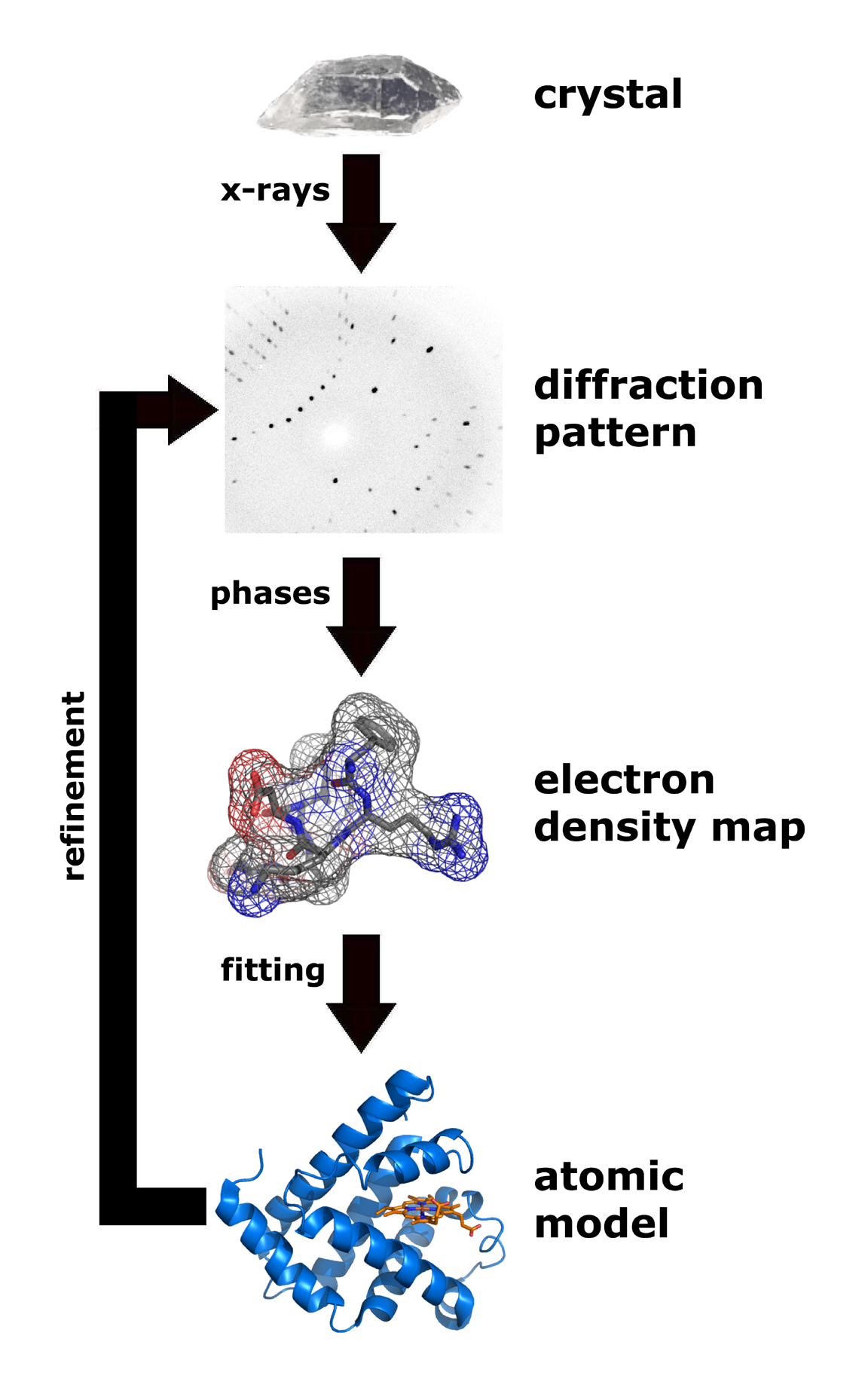
X-ray Diffraction: Solving the Crystal Structure of Molecules
X-ray diffraction (XRD) is the gold standard for determining the precise atomic structure of crystalline substances. When X-rays are directed at a crystal, they scatter in specific patterns based on the crystal’s atomic arrangement. By analyzing these patterns, scientists can create detailed models of molecular structures.
How X-ray Diffraction Works
X-rays interact with electrons in the crystal, producing a diffraction pattern based on the atomic arrangement. By measuring the angles and intensities of these scattered rays, researchers can reconstruct the crystal’s three-dimensional structure.
 |
| Attribution: Thomas Splettstoesser (www.scistyle.com), CC BY-SA 3.0 <https://creativecommons.org/licenses/by-sa/3.0>, via Wikimedia Commons |
Applications of X-ray Diffraction
X-ray diffraction has been crucial in structural biology and drug design. It’s used to reveal the structures of complex molecules, like proteins and DNA, providing insights that help scientists understand how these molecules work. XRD has also been essential in developing new drugs by revealing the structure of molecular targets.
Light Scattering: Studying Molecular Size and Interactions
Light scattering techniques provide information about the size, shape, and interaction of molecules in solution. By observing how light scatters off molecules, scientists can gather details about molecular weight, particle size, and interactions.
How Light Scattering Works
When light hits molecules or particles, it scatters in different directions. By analyzing the scattered light, researchers can determine molecular size, shape, and interaction patterns without needing to separate the molecules.
Applications of Light Scattering
Light scattering is used to study macromolecules like proteins and polymers. It’s also common in nanoparticle research and drug formulation, allowing researchers to analyze particles and understand their stability in complex mixtures.
Mass Spectrometry: Identifying Molecules with Precision
Mass spectrometry (MS) is an incredibly sensitive method for identifying and analyzing the mass and composition of molecules. MS is used to detect trace amounts of compounds, analyze complex mixtures, and identify unknown substances with precision.
 |
| Attribution- SCB2024, CC0, via Wikimedia Commons |
How Mass Spectrometry Works
In MS, molecules are ionized, and the ions are sorted based on their mass-to-charge ratio. This generates a spectrum that acts like a “fingerprint” for each compound. Different types of mass spectrometry—such as Matrix-Assisted Laser Desorption/Ionization (MALDI) and Electrospray Ionization (ESI)—make it possible to analyze a wide range of molecules.
 |
| Attribution: Philippe Hupé, CC BY-SA 3.0 <https://creativecommons.org/licenses/by-sa/3.0>, via Wikimedia Commons |
 |
| Attribution: OmarKana, CC BY-SA 4.0 <https://creativecommons.org/licenses/by-sa/4.0>, via Wikimedia Commons |
Applications of Mass Spectrometry
Mass spectrometry is a cornerstone in fields like proteomics, drug discovery, and environmental science. It can identify molecules, quantify compounds, and study protein modifications, providing crucial information for biological and chemical research.
Surface Plasmon Resonance (SPR): Watching Molecules Interact in Real Time
Surface Plasmon Resonance (SPR) is a powerful tool for studying molecular interactions in real time without labeling. SPR measures changes in refractive index near a sensor surface, allowing scientists to observe binding events between molecules.
.jpg) |
| Attribution: SariSabban, CC BY-SA 3.0 <https://creativecommons.org/licenses/by-sa/3.0>, via Wikimedia Commons |
How SPR Works
One molecule (such as an antibody) is attached to a sensor chip, and a sample containing a potential binding partner flows over it. As binding occurs, the refractive index near the sensor surface changes, creating a binding curve that shows the interaction’s strength and rate.
_Operations_A.jpg) |
| Fig- The diagram summarizes the data displayed by a Surface Plasmon Resonance (SPR) machine using a single ligand. Attribution: SariSabban, CC BY-SA 3.0 <https://creativecommons.org/licenses/by-sa/3.0>, via Wikimedia Commons |
_Operations_B.jpg) |
| Fig.-Diagram summarizes the data displayed by a Surface Plasmon Resonance (SPR) machine using a capturing molecule followed by a ligand Attribution: SariSabban, CC BY-SA 3.0 <https://creativecommons.org/licenses/by-sa/3.0>, via Wikimedia Commons |
Applications of SPR
SPR is widely used in drug development, immunology, and molecular biology to study how molecules bind to one another. It’s invaluable for screening drug candidates, studying biomolecular interactions, and investigating binding affinities.
Conclusion: The Marvels of Biophysical Methods in Modern Science
Today’s biophysical methods are like windows into the molecular world, giving us the tools to analyze and understand life’s building blocks at a level of detail that was once unimaginable. From UV/visible spectroscopy to mass spectrometry and surface plasmon resonance, each method offers unique insights, helping scientists uncover the mysteries of molecular structure, function, and interaction.
These techniques aren’t just tools for science—they’re essential for progress in fields as varied as medicine, environmental science, and materials research. As technology continues to evolve, biophysical methods will keep improving, providing us with even deeper insights into the molecular world. These insights will continue to fuel breakthroughs in drug discovery, disease research, and the development of new materials, ultimately shaping the future of science and medicine.
Whether we’re studying DNA structures, tracking protein interactions, or analyzing tiny nanoparticles, biophysical methods provide the precision and depth that modern research demands. In a world where even the smallest details matter, these methods are essential for unlocking the secrets of life and driving scientific discovery forward.